

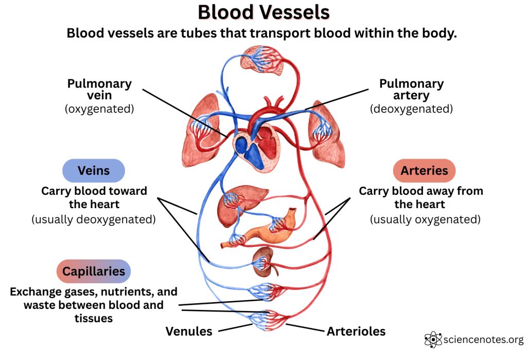
Blood vessels are tubes that transport blood throughout the body. Together with the heart, they make up the circulatory system. Blood vessels deliver oxygen, nutrients, hormones, and immune cells to tissues while also removing waste products like carbon dioxide and urea. They play a critical role in maintaining homeostasis, regulating body temperature, and ensuring tissue viability.
Key Points About Blood Vessels
- Blood vessels are tubes that transport blood.
- There are three main types of blood vessels: arteries, veins, and capillaries.
- Arteries carry blood away from the heart; veins return it to the heart; capillaries allow exchange with tissues.
- The human body contains around 60,000 miles (96,000 km) of blood vessels.
- Blood vessel walls have three layers, but these layers differ in structure between arteries, veins, and capillaries.
- Healthy blood vessels are flexible and resilient, while diseased vessels become stiff, narrowed, or blocked.
- Disorders of blood vessels contribute to conditions such as hypertension, stroke, and aneurysms.
Types of Blood Vessels
Blood vessels fall into several categories based on their size, function, and direction of blood flow. Each type plays a unique role in circulating blood throughout the body. The three main types of blood vessels are arteries, veins, and capillaries. However, there are intermediate-size vessels and other subcategories.
1. Arteries
- Function: Carry oxygenated blood away from the heart (except pulmonary arteries, which carry deoxygenated blood to the lungs).
- Location: Found deep within the body, often near bones.
- Structure: Thick, muscular, and elastic to handle high pressure from the heart.
- Examples: Aorta, carotid arteries, femoral arteries.
Types of Arteries
Arteries are classified by size and function:
| Type | Description | Examples |
|---|---|---|
| Elastic arteries | Large arteries closest to the heart; contain more elastic tissue to withstand high pressure from ventricular contraction. | Aorta, pulmonary trunk |
| Muscular arteries | Medium-sized arteries with thick smooth muscle layers to distribute blood and control flow. | Brachial, femoral, radial arteries |
| Arterioles | Smallest arteries that lead into capillary beds; critical for regulating blood pressure. | Arterioles throughout organs |
- Note: The elastic recoil of elastic arteries helps maintain blood pressure between heartbeats, while muscular arteries help direct blood to specific tissues.
2. Arterioles
- Function: Small branches of arteries that regulate blood flow into capillaries.
- Location: Connect arteries to capillary beds.
- Structure: Smaller diameter with muscular walls for controlling blood pressure.
3. Capillaries
- Function: Allow the exchange of gases, nutrients, and waste between blood and tissues.
- Location: Penetrate nearly all body tissues.
- Structure: One cell layer thick (endothelium) to permit diffusion.
Types of Capillaries
Capillaries are not all the same. There are three major types, each specialized for different levels of permeability:
| Type | Structure and Function | Found In |
|---|---|---|
| Continuous | Most common; endothelial cells form a tight, uninterrupted lining. | Skin, muscles, lungs, brain |
| Fenestrated | Have small pores (fenestrations) that allow faster exchange of small molecules. | Kidneys, intestines, endocrine glands |
| Sinusoidal | Have large gaps between cells for movement of larger molecules and cells. | Liver, bone marrow, spleen |
Capillary Beds and Regulation
Capillaries are organized into capillary beds, networks controlled by precapillary sphincters that open and close to regulate blood flow based on tissue needs. This regulation is essential for thermoregulation and nutrient distribution.
4. Venules
- Function: Collect blood from capillaries and begin the return to the heart.
- Location: Between capillaries and veins.
- Structure: Thin walls, some with valves.
5. Veins
- Function: Carry deoxygenated blood back to the heart (except pulmonary veins, which carry oxygenated blood from the lungs).
- Location: Closer to the surface of the skin, often visible.
- Structure: Thinner walls than arteries, with valves to prevent backflow.
Types of Veins
Veins vary by size and location in the body:
| Type | Description | Examples |
|---|---|---|
| Large veins | Carry blood directly into the heart; have thin walls and wide lumens. | Superior/inferior vena cava |
| Medium veins | Collect blood from limbs and organs; contain valves to prevent backflow. | Brachial, saphenous, jugular veins |
| Venules | Smallest veins that receive blood from capillaries. | Venules in all tissue beds |
| Superficial veins | Located near the surface of the body; often visible under the skin. | Cephalic, basilic veins |
| Deep veins | Accompany arteries and lie deeper within muscles. | Femoral, popliteal veins |
Anatomy of Blood Vessels

Although blood vessels differ in function, many share a common layered structure. Understanding the anatomy of blood vessels reveals how they maintain pressure, regulate flow, and interact with surrounding tissues.
Most blood vessels (except capillaries) consist of three layers:
| Layer | Description | Found In |
|---|---|---|
| Tunica intima | Innermost layer made of endothelium; smooth to reduce friction | Arteries, veins |
| Tunica media | Middle layer of smooth muscle and elastic tissue | Thick in arteries |
| Tunica externa (adventitia) | Outermost layer of connective tissue | Thicker in veins |
Comparisons
- Arteries: Thick tunica media, narrow lumen, no valves.
- Veins: Thinner walls, larger lumen, contain valves.
- Capillaries: Only have tunica intima (a single layer of endothelial cells).
Mnemonics and Quiz
Here are some helpful tricks for remembering blood vessel facts:
Mnemonics
- “Arteries Away” – Arteries carry blood away from the heart.
- “LAV: Lumen-Artery-Vein” – Arteries have a Larger tunica media and smaller lumen than veins.
- “Very Tiny Capillaries” – The order of decreasing vessel diameter: Vein → Venule → Capillary.
- “CAVe” for blood flow direction:
- Capillaries
- Arteries
- Veins – helps track where blood goes during systemic circulation.
Blood Vessels and Circulation Quiz
Appearance of Blood Vessels
Blood vessels vary in size, shape, and visibility.
- Arteries: Round, thick-walled, usually deep red when oxygenated.
- Veins: Flattened or collapsed appearance, bluish through the skin due to light scattering (deoxygenated blood is still red).
- Capillaries: Microscopic and invisible to the naked eye; form dense networks within tissues.
How Blood Vessels Work in Circulation
Blood vessels form a closed-loop system that transports blood in a continuous circuit. Here’s a step-by-step walkthrough of how this system works in humans, starting and ending at the heart:
1. Oxygen-Poor Blood Enters the Right Side of the Heart
- From: The superior and inferior vena cava (large veins from the body)
- To: The right atrium, then pumped into the right ventricle
2. Blood Goes to the Lungs
- The pulmonary arteries carry deoxygenated blood from the right ventricle to the lungs.
- In the pulmonary capillaries, carbon dioxide is released and oxygen is absorbed.
3. Oxygen-Rich Blood Returns to the Heart
- The pulmonary veins return oxygenated blood to the left atrium.
- Blood then moves into the left ventricle, the heart’s most powerful chamber.
4. Blood Is Pumped Into the Body
- The aorta, the body’s largest artery, carries oxygen-rich blood to all tissues.
- Blood travels through elastic arteries, then muscular arteries, and finally arterioles.
5. Exchange Occurs in the Capillaries
- In the systemic capillaries, oxygen and nutrients diffuse into tissues.
- Waste products like carbon dioxide diffuse into the blood.
6. Blood Returns via the Veins
- Blood from capillaries drains into venules, then into medium and large veins.
- Valves in the veins prevent backflow and assist with return to the heart, especially from the lower body.
7. Cycle Repeats
- The deoxygenated blood enters the heart’s right atrium, completing the loop and beginning the cycle again.
Symptoms of Blood Vessel Problems
Problems with blood vessels produce a wide range of symptoms. Recognizing early signs may help prevent serious complications such as stroke or cardiovascular disease.
Symptoms vary by the vessel type and disorder but may include:
- Pain or cramping in the limbs (claudication)
- Cold or pale extremities
- Swelling or edema
- Visible varicose veins
- Dizziness or fainting
- Headaches or blurred vision (in hypertension)
- Ulcers or slow-healing wounds
- Chest pain or shortness of breath (if affecting coronary vessels)
Blood Vessel Diseases and Disorders
Numerous conditions affect the structure or function of blood vessels. Some are acute and life-threatening, while others develop slowly over time and require long-term management.
Common Conditions
| Disorder | Description |
|---|---|
| Atherosclerosis | Fatty plaque buildup in arteries, reducing blood flow |
| Aneurysm | Bulging or weakened section of an artery wall |
| Varicose veins | Enlarged, twisted veins due to valve failure |
| Deep vein thrombosis (DVT) | Blood clot in a deep vein, often in the leg |
| Raynaud’s disease | Narrowing of small arteries in response to cold or stress |
| Vasculitis | Inflammation of blood vessels |
| Hypertension | Chronic high blood pressure, straining vessel walls |
| Stroke | Interruption of blood supply to the brain |
| Peripheral artery disease (PAD) | Narrowing of arteries in limbs |
Diagnosis of Blood Vessel Disorders
Diagnosing vascular conditions often involves a combination of physical exams, imaging techniques, and blood tests. Early detection improves treatment outcomes.
Doctors use several tools to diagnose vascular problems:
- Physical exam (checking pulses, skin color, temperature)
- Blood pressure measurement
- Ultrasound (especially Doppler imaging for flow)
- Angiography (X-ray with dye to view vessels)
- CT or MRI scans
- Blood tests (e.g., for clotting factors or cholesterol)
- Ankle-brachial index (for PAD detection)
Maintaining Healthy Blood Vessels
Healthy lifestyle choices significantly reduce the risk of blood vessel problems. This section outlines evidence-based habits for keeping your vascular system in top condition.
- Exercise regularly to promote circulation and vessel elasticity.
- Eat a heart-healthy diet: low in saturated fat, salt, and sugar; rich in fiber and antioxidants.
- Avoid smoking, which damages endothelial cells and promotes plaque buildup.
- Maintain a healthy weight to reduce strain on the vascular system.
- Control blood pressure, cholesterol, and blood sugar through lifestyle or medications.
- Stay hydrated to keep blood flowing smoothly.
- Manage stress, which can contribute to vessel constriction and high blood pressure.
Frequently Asked Questions (FAQs)
What are the smallest blood vessels in the body?
Capillaries are the smallest blood vessels. They are only one cell thick and are involved in the exchange of gases and nutrients.
Why do veins look blue under the skin?
Veins appear blue because of the way light penetrates the skin and is absorbed or scattered. The blood in veins is dark red, but the skin and tissues filter out red wavelengths.
Can arteries carry deoxygenated blood?
Yes, pulmonary arteries carry deoxygenated blood from the heart to the lungs, which is an exception to the general rule. Similarly the pulmonary veins carry oxygenated blood, making them the other exception.
How many blood vessels are there?
The human body contains an estimated 60,000 to 100,000 miles (96,000 to 160,000 kilometers) of blood vessels. This includes arteries, veins, and capillaries.
What causes varicose veins?
They result from weakened or damaged valves in veins, leading to blood pooling and vein enlargement.
How can I improve poor circulation?
Regular physical activity, elevating legs, avoiding sitting or standing for long periods, and wearing compression garments all help improve circulation.
What is the difference between an artery and a vein?
Arteries carry blood away from the heart and are under higher pressure, with thicker muscular walls. Veins carry blood back to the heart, have valves, and are under lower pressure.
Fun Facts About Blood Vessels
- Your blood vessels could circle the Earth: If laid end to end, the blood vessels in your body would stretch 60,000 to 100,000 miles—enough to wrap around the Earth more than twice.
- The aorta is the largest blood vessel: It is roughly 2.5 cm (1 inch) in diameter, about the size of a garden hose.
- Capillaries are tiny but numerous: The majority of blood vessels in the body are capillaries, with billions forming dense networks in nearly every tissue.
- Veins contain most of your blood: At any given moment, about 70% of your blood is in your veins.
- Pulse points are arteries: The throbbing you feel at your wrist or neck is an artery expanding with each heartbeat.
- Varicose veins aren’t just cosmetic: Sometimes they signal underlying valve problems that impair blood return to the heart.
- Blood flows fastest in arteries and slowest in capillaries: This slow flow in capillaries allows efficient exchange of gases and nutrients with tissues.
References
- Erbel, R.; Eggebrecht, H. (2006). “Aortic dimensions and the risk of dissection”. Heart. 92 (1): 137–142. doi:10.1136/hrt.2004.055111
- Poole, D.C.; Kano, Y.; Koga, S.; et al. (2021). “August Krogh: Muscle capillary function and oxygen delivery”. Comparative Biochemistry and Physiology. Part A, Molecular & Integrative Physiology. 253: 110852. doi:10.1016/j.cbpa.2020.110852
- Prisby, R.D. (2017). “Mechanical, hormonal and metabolic influences on blood vessels, blood flow and bone”. The Journal of Endocrinology. 235 (3): R77 – R100. doi:10.1530/JOE-16-0666
- Tucker, W.D.; Arora, Y.; Mahajan, K. [2023] Anatomy, Blood Vessels. NIH National Library of Medicine. Available from: https://www.ncbi.nlm.nih.gov/books/NBK470401/
- Qin G, Wang L, Hua Y, et al. (2020). “Comparative morphology of the internal elastic lamina of cerebral and peripheral arteries“. International Journal of Clinical and Experimental Pathology. 13 (4): 764–770.