
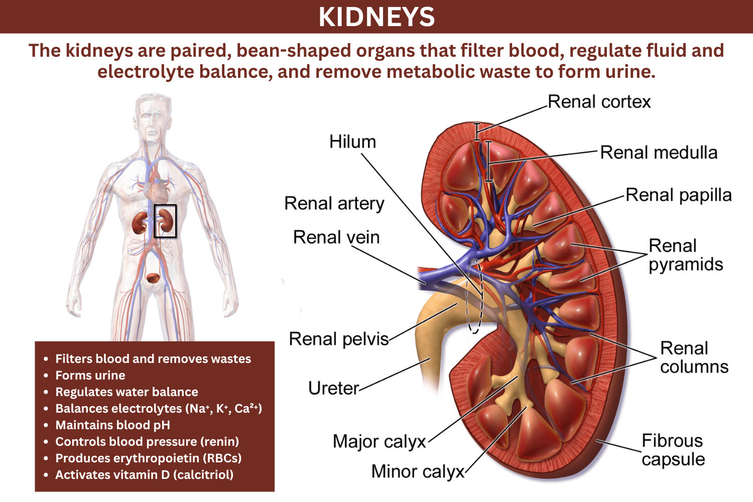
The kidneys are paired, bean-shaped organs that filter blood, regulate fluid and electrolyte balance, and remove metabolic waste to form urine. These organs are essential for maintaining homeostasis, ensuring that the internal environment of the body remains stable despite changes in diet, activity, and external conditions. In addition to filtration, the kidneys act as endocrine organs, influencing red blood cell production, calcium metabolism, and blood pressure.
Key Takeaways: Kidneys
- Kidneys are paired retroperitoneal organs that filter blood and produce urine.
- Each kidney contains about 1 million nephrons, the functional filtration units.
- They regulate water balance, electrolytes, blood pressure, and pH.
- Kidneys produce hormones, including erythropoietin and renin.
- Renal blood flow is high, about 20–25% of cardiac output.
- Damage to kidneys can lead to fluid imbalance, toxin buildup, and systemic disease.
What Are the Kidneys?
The kidneys are vital organs of the urinary system responsible for filtering blood plasma, removing waste products (such as urea, creatinine, and excess ions), and maintaining the chemical composition of body fluids. Every minute, large volumes of blood pass through the kidneys, where selective filtration and reabsorption ensure that essential substances such as glucose, amino acids, and water remain in circulation.
The kidneys are not passive filters. They actively monitor and adjust blood composition in response to physiological needs. For example, they conserve water during dehydration, excrete excess ions after a salty meal, and adjust acid–base balance during metabolic disturbances.
Human Kidneys: Location, Number, Size, and Appearance
Humans typically have two kidneys located on either side of the vertebral column in the upper posterior abdomen.
- Location: Retroperitoneal, on either side of the vertebral column (T12–L3)
- Position difference: The right kidney sits slightly lower due to the liver
- Number: Usually two, although individuals can live with one
- Size: About 10–12 cm long, 5–7 cm wide, 2–3 cm thick
- Weight: Approximately 120–150 g each in adults
- Appearance: Smooth, reddish-brown, bean-shaped with a concave medial border
The concave side of each kidney faces inward and contains the hilum, where blood vessels, nerves, and the ureter enter or exit.
Differences Between Males and Females
- Male kidneys are typically slightly larger due to greater average body mass
- Hormonal differences influence renal physiology, particularly fluid handling and blood pressure regulation
- Structural differences are minimal, and both sexes have the same basic anatomy
Structure of the Kidneys
Each kidney is surrounded by protective and supportive layers that help maintain its position and protect it from injury.
- Renal capsule: A thin, tough fibrous layer that directly surrounds the kidney
- Perirenal fat: A cushioning layer of adipose tissue that absorbs shock
- Renal fascia: A connective tissue layer that anchors the kidney to surrounding structures
Internally, the kidney is organized into two main regions:
- Renal cortex: The outer region, rich in blood vessels and filtration structures
- Renal medulla: The inner region, organized into pyramids that concentrate urine
This structural organization reflects the functional division of labor between filtration and urine concentration.
Gross Anatomy of the Kidneys
The gross anatomy describes the structures visible without a microscope and reveals how urine flows through the kidney.
- Renal cortex: Contains glomeruli and convoluted tubules where filtration begins
- Renal medulla: Composed of renal pyramids that direct urine inward
- Renal pyramids: Cone-shaped structures containing loops of Henle and collecting ducts
- Renal papilla: The tip of each pyramid where urine drains
- Minor and major calyces: Channels that collect urine from papillae
- Renal pelvis: A funnel-shaped chamber that leads to the ureter
- Hilum: The medial indentation where vessels, nerves, and ureter pass
Urine flows in a continuous pathway from the cortex through the medulla and into the renal pelvis before entering the ureter.
Kidney Microanatomy

The nephron is the microscopic functional unit responsible for filtration and urine formation. Each kidney contains approximately one million nephrons.
Components of a Nephron
- Renal corpuscle:
- Glomerulus (a network of capillaries)
- Bowman’s capsule (a surrounding structure that collects filtrate)
- Renal tubule:
- Proximal convoluted tubule (PCT)
- Loop of Henle
- Distal convoluted tubule (DCT)
- Collecting duct system
Functional Processes
- Filtration: Blood plasma is filtered under pressure in the glomerulus
- Reabsorption: Essential substances such as glucose, ions, and water are returned to the bloodstream
- Secretion: Additional wastes and excess ions are actively transported into the filtrate
The arrangement of nephrons allows for fine control of fluid composition, making the kidneys highly efficient regulators of internal balance.
Kidney vs Nephron vs Urinary System
Understanding how these terms relate helps clarify kidney function.
- Kidney: The organ responsible for filtering blood
- Nephron: The microscopic functional unit within the kidney
- Urinary system: Includes kidneys, ureters, bladder, and urethra
Each level represents a different scale of organization, from microscopic to whole-system function.
Renal Blood Supply
The kidneys receive a remarkably high blood flow relative to their size, reflecting their filtration role.
- Renal artery → segmental arteries → interlobar arteries → arcuate arteries → cortical radiate arteries
- Blood enters the afferent arteriole, then flows into the glomerulus
- It exits via the efferent arteriole, forming:
- Peritubular capillaries (associated with cortical nephrons)
- Vasa recta (associated with juxtamedullary nephrons)
This dual capillary system is unique and allows for both filtration and precise reabsorption.
Nerve Supply
Kidneys receive autonomic innervation that regulates their function.
- Sympathetic innervation: Controls renal blood flow and stimulates renin release
- Parasympathetic input: Limited influence
- Sensory fibers convey pain signals, often perceived as flank or lower back pain
Kidney Development
Kidneys develop from intermediate mesoderm through three sequential stages:
- Pronephros: Early, nonfunctional structure
- Mesonephros: Temporary functioning kidney during early development
- Metanephros: The permanent kidney
The metanephros begins forming around the fifth week of gestation. Interaction between the ureteric bud and metanephric mesenchyme drives the formation of nephrons and collecting ducts.
How the Kidneys Work
Kidneys continuously filter blood through a coordinated sequence of processes that produce urine while maintaining internal balance.
Blood enters the kidney through the renal artery and is delivered to nephrons. In each nephron, the glomerulus acts as a high-pressure filter, forcing water and small solutes into Bowman’s capsule while retaining cells and large proteins.
The resulting filtrate travels through the renal tubule, where its composition is carefully adjusted:
- In the proximal convoluted tubule, most water, glucose, and ions are reabsorbed
- In the loop of Henle, a countercurrent system establishes a concentration gradient in the medulla, allowing water conservation
- In the distal convoluted tubule and collecting duct, hormones such as aldosterone and antidiuretic hormone regulate final ion and water balance
By the time the fluid reaches the collecting ducts, it has been transformed into urine. The kidneys produce about 1–2 liters of urine per day from roughly 150–180 liters of initial filtrate.
Renal Physiology: Filtration Rate and Regulation
Kidney function depends on the rate at which blood is filtered, known as the glomerular filtration rate (GFR). GFR reflects how much filtrate forms per minute across all nephrons and serves as a key indicator of kidney health.
- Normal GFR: Approximately 90–120 mL/min/1.73 m² in healthy adults
- Filtration pressure: Determined by the balance of hydrostatic and osmotic forces in the glomerulus
- Clinical relevance: Declining GFR indicates impaired kidney function
Several factors influence GFR:
- Blood pressure: Higher pressure increases filtration, within limits
- Afferent arteriole diameter: Dilation increases GFR, constriction decreases it
- Efferent arteriole diameter: Moderate constriction increases GFR, but excessive constriction reduces blood flow
- Plasma protein concentration: Higher protein levels reduce filtration due to osmotic pressure
The kidneys maintain relatively stable GFR through autoregulation:
- Myogenic response: Smooth muscle in afferent arterioles constricts or dilates in response to pressure changes
- Tubuloglomerular feedback: The macula densa senses sodium concentration and adjusts afferent arteriole tone
Clinically, GFR is estimated as eGFR using blood creatinine levels, age, sex, and other factors.
Functions of the Kidneys
Kidneys perform multiple essential physiological roles.
Filtration and Waste Removal
- Remove nitrogenous wastes such as urea and creatinine
- Eliminate toxins, drugs, and metabolic byproducts
Fluid and Electrolyte Balance
- Regulate sodium, potassium, calcium, and water levels
Acid–Base Balance
- Maintain blood pH by excreting hydrogen ions and conserving bicarbonate
Blood Pressure Regulation
- Control blood volume and release renin
Hormone Production
- Erythropoietin: Stimulates red blood cell production
- Calcitriol: Active vitamin D for calcium regulation
Hormonal Regulation of Kidney Function
Kidney function is tightly regulated by hormones that coordinate fluid balance, blood pressure, and electrolyte levels.
- Renin–angiotensin–aldosterone system (RAAS):
Triggered by low blood pressure or low sodium
Renin release leads to angiotensin II formation, causing vasoconstriction and aldosterone release - Aldosterone:
Increases sodium reabsorption and potassium excretion in the distal nephron
Water follows sodium, increasing blood volume - Antidiuretic hormone (ADH):
Increases water permeability in the collecting ducts
Promotes water reabsorption and concentrates urine - Atrial natriuretic peptide (ANP):
Released by the heart in response to high blood volume
Promotes sodium and water excretion, lowering blood pressure
These hormonal systems allow the kidneys to respond dynamically to changes in hydration and circulation.
Countercurrent Mechanism and Urine Concentration
The kidneys produce either dilute or highly concentrated urine, depending on the body’s needs. This ability depends on the countercurrent mechanism in the nephron.
- Countercurrent multiplier (loop of Henle):
Establishes a concentration gradient in the renal medulla
Descending limb is permeable to water, while ascending limb actively transports ions - Countercurrent exchanger (vasa recta):
Preserves the gradient by exchanging solutes and water with surrounding tissue - Medullary gradient:
Allows water to be reabsorbed from collecting ducts when ADH is present
This system enables efficient water conservation. Animals adapted to dry environments often have longer loops of Henle, which enhance urine concentration.
Kidney vs Other Excretory Organs
The kidneys are the primary excretory organs, but other systems also contribute to waste removal.
- Kidneys: Remove nitrogenous wastes, regulate ions, control water balance
- Lungs: Remove carbon dioxide and help regulate pH
- Skin: Excretes small amounts of water, salts, and urea through sweat
- Liver: Detoxifies chemicals and converts ammonia into urea
Together, these organs maintain internal balance by removing different types of waste products.
Clinical Significance of the Kidneys
Kidney disorders affect nearly every system in the body because of their central role in homeostasis.
- Acute kidney injury (AKI): Sudden decline in function
- Chronic kidney disease (CKD): Progressive loss of filtration capacity
- Kidney stones: Crystalline deposits that obstruct urine flow
- Glomerulonephritis: Inflammation of filtering structures
- Polycystic kidney disease: Genetic disorder causing cyst formation
Advanced kidney failure requires dialysis or transplantation.
Symptoms of Kidney Dysfunction
Kidney disease often develops silently, but common symptoms include:
- Swelling (edema), especially in legs and around the eyes
- Fatigue and weakness
- Changes in urination, including frequency, color, or foaminess
- High blood pressure
- Nausea or decreased appetite
- Persistent flank or lower back pain
Laboratory Tests and Kidney Function Assessment
Doctors evaluate kidney function using blood and urine tests.
- Serum creatinine:
A waste product from muscle metabolism
Elevated levels indicate reduced kidney function - Blood urea nitrogen (BUN):
Measures urea concentration in blood
Can increase with kidney dysfunction or dehydration - Estimated GFR (eGFR):
Calculated from creatinine levels
Used to stage chronic kidney disease - Urinalysis:
Detects abnormalities such as protein, glucose, or blood
Provides insight into kidney damage or infection
These tests allow early detection and monitoring of kidney disease.
Kidney Stones: Types and Formation
Kidney stones form when dissolved minerals crystallize in urine.
- Calcium oxalate stones: Most common type
- Uric acid stones: Associated with high purine intake or acidic urine
- Struvite stones: Often linked to urinary tract infections
- Cystine stones: Caused by a rare genetic disorder
Risk factors include dehydration, diet, genetics, and metabolic conditions. Preventive strategies focus on hydration and dietary management.
Dialysis and Kidney Transplantation
When kidneys fail, medical interventions can replace some of their functions.
- Hemodialysis:
Blood is filtered through an external machine
Removes waste and excess fluid - Peritoneal dialysis:
Uses the peritoneal membrane as a natural filter
Fluid exchanges occur within the abdominal cavity - Kidney transplantation:
Surgical replacement with a donor kidney
Requires lifelong immunosuppressive therapy
These treatments sustain life but do not fully replicate all kidney functions.
Maintaining Kidney Health
Healthy kidneys depend on lifestyle and preventive care.
- Stay well hydrated
- Maintain healthy blood pressure and blood sugar
- Limit excessive salt intake
- Avoid overuse of NSAIDs and other nephrotoxic drugs
- Eat a balanced diet rich in fruits and vegetables
- Exercise regularly
- Get routine medical checkups
Evolutionary Adaptations of Kidneys
Kidneys have evolved to meet environmental challenges.
- Freshwater animals: Excrete dilute urine to eliminate excess water
- Marine animals: Conserve water and excrete excess salts
- Desert mammals: Produce highly concentrated urine
- Nitrogen waste forms:
- Ammonia (aquatic species)
- Urea (mammals)
- Uric acid (birds and reptiles)
These adaptations reflect differences in habitat and water availability.
Kidneys in Other Vertebrates
Kidneys vary among vertebrates based on environmental and physiological demands.
- Fish: Regulate salt and water in aquatic environments
- Amphibians: Adapt to both aquatic and terrestrial life
- Reptiles and birds: Excrete uric acid to conserve water
- Mammals: Possess loops of Henle that enable urine concentration
Desert mammals often have especially long loops of Henle, allowing them to produce highly concentrated urine and conserve water efficiently.
Common Misconceptions
- “Kidneys only remove waste.”
They also regulate hormones, blood pressure, and electrolyte balance. - “You need two kidneys to survive.”
One kidney can perform all necessary functions. - “Kidney disease always causes pain.”
Many kidney diseases are asymptomatic in early stages. - “Drinking more water always improves kidney function.”
Excessive water intake can disrupt electrolyte balance.
FAQs
What is the main function of the kidneys?
To filter blood, remove waste, and regulate fluid and electrolyte balance.
Can a person live with one kidney?
Yes, one kidney can compensate for the loss of the other.
How much blood do kidneys filter daily?
About 150–180 liters of filtrate are produced daily, with most reabsorbed.
What causes kidney stones?
Crystallization of minerals such as calcium oxalate in urine.
How do kidneys regulate blood pressure?
By controlling fluid volume and releasing renin.
Are kidney problems reversible?
Some acute conditions are reversible, but chronic disease is often progressive.
Can you live without a kidney?
A person can live with one kidney, but not without any functioning kidney tissue. Without kidney function, dialysis or a transplant is required.
References and Further Reading
- Alpern, Robert J.; Caplan, Michael; Moe, Orson W. (2012). Seldin and Giebisch’s The Kidney: Physiology and Pathophysiology. Academic Press. ISBN 978-0-12-381463-0.
- Bard, J.; Vize, P.D.; Woolf, A.S. (2003). The Kidney: from normal development to congenital disease. Boston: Academic Press.ISBN 978-0-12-722441-1.
- Barrett, K.E.; Barman, S.M.; Yuan, J.X.; Brooks, H. (2019). Ganong’s Review of Medical Physiology (26th ed.). New York. ISBN 978-1-260-12240-4.
- Lv, J.C.; Zhang, L.X. (2019). “Prevalence and Disease Burden of Chronic Kidney Disease”. Renal Fibrosis: Mechanisms and Therapies. Advances in Experimental Medicine and Biology. 1165: 3–15. doi:10.1007/978-981-13-8871-2_1. ISBN 978-981-13-8871-2.
- Zhou, Xin J.; Laszik, Zoltan G.; Nadasdy, Tibor; D’Agati, Vivette D. (2017). Silva’s Diagnostic Renal Pathology. Cambridge University Press. ISBN 978-1-316-61398-6.